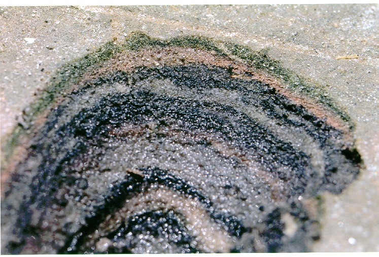
Farbstreifen-Sandwatt
Die nachfolgenden Bilder und Texte stammen aus einem Projekt des Sommerkurses Microbial Diversity am Marine Biological Laboratory in Woods Hole, MA, USA. Die Urheberrechte der Bilder liegen bei Elke Jaspers und Rolf Schauder.
Microbial Diversity Course 1997, MBL, Woods Hole
Microbial Mats
Take a knife and cut into the firm sand in a salt marsh. This is what you may find: Colored bands in the sand. In the upper 2 to 5 mm you'll find a green and a pink layer. The green layer contains diatoms and cyanobacteria. The color of the pink layer stems from carotinoids, the major pigments of phototrophic purple sulfur bacteria. Just a little bit deeper you'll find a black layer - iron sulfide, precipitated from iron ions with sulfide formed by anoxic sulfate reducing bacteria. These bacteria decompose the organic matter formed in the top layers, and thus recycle the biomass. Underneath the colored bands the sand turns grey due to a chemical formation of pyrite. Below that you'll find more layers - left-overs from passed years. The hole in the sand has a size of about 5 cm (2 inch).
Some of the bacteria of the so called "Farbstreifen-Sandwatt" are presented here on this page. The samples were taken at the Great Sippewissett Saltmarsh at Woods Hole, MA, USA.
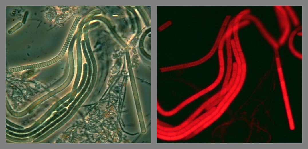
Green layer
Filamentous cyanobacteria from the top (green) layer of a microbial mat in the Great Sippewisset Saltmarsh. Left: phase contrast, right: The same view after illumination with green light. The red autoflourescence demonstrates the presence of phycobiliproteins
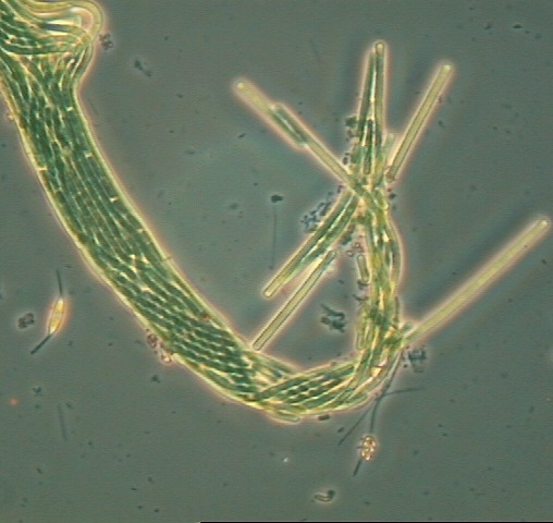
Green layer
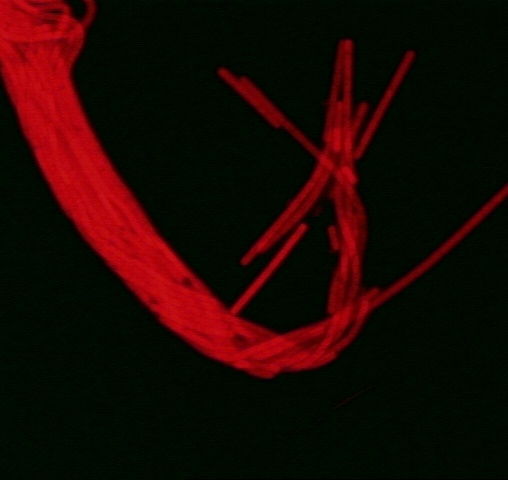
Some more filamentous cyanobacteria from the green layer of a microbial mat from the Great Sippewissett Saltmarsh. Left: phase contrast micrograph, right: autofluorescence of the same view after illumination with green light. Length of the borderline: approx. 0.4 mm
Green layer
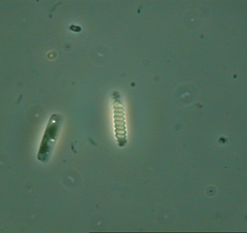
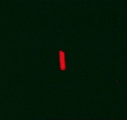
A short fragment of the cyanobacterium Spirulina spec. (center) and a diatom. Left: phase contrast. Right: Autofluorescence of the same view after illumination with green light. The weak fluorescence of the diatom due to its chlorophyll a cannot be seen. Diameter of the microbes: approx: 3 µm
Green layer
Lyngbya spec, a sheeted cyanobacteriam found in the Great Sippewissett Saltmarsh. Two cells are leaving the sheet. Left: phase contrast micrograph. right: autofluorescence after illumination with green light.
Size of the image: 100X100 µm
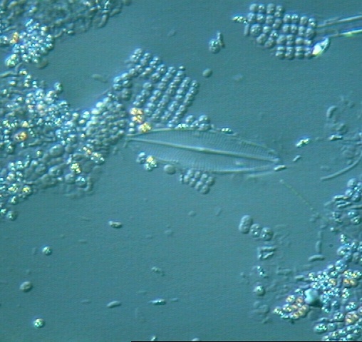
Pink layer
The major microbes of the pink layer are phototrophic purple sulfur bacteria. The egg shaped organisms are diatoms. Phase contrast.
75x75 µm
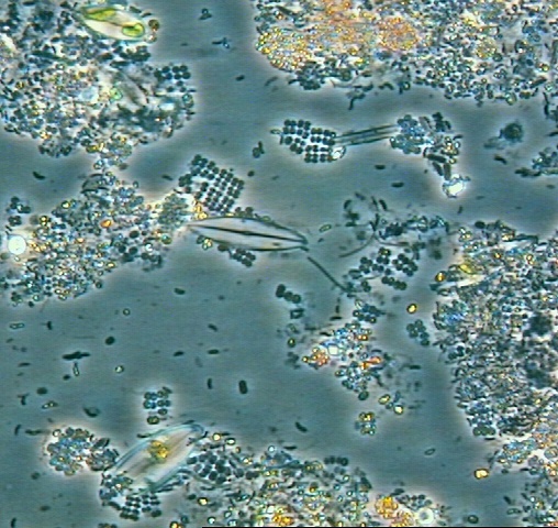
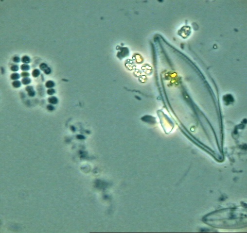
Pink layer
Purple sulfur bacteria and a diatom from the pink layer of a microbial mat from the Great Sippewissett Saltmarsh. The bright spots in the cells are sulfur granules. DIC micrograph.
75x75 µm
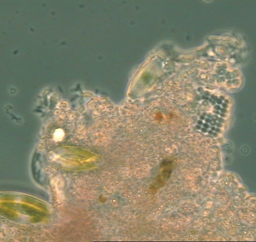
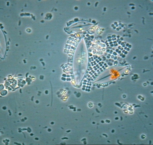
Pink layer
A typical bright field micrograph of microbes of the pink layer of microbial mats. The layer contains various purple sulfur bacteria (mostly in packets), some diatoms (center), and some single cells.
Size of the picture: 150x150 µm
Pink layer
Close-up of a group of purple sulfur bacteria (Thiopedia spec) clustered around a cell of a diatom.
Phase contrast
Size of the picture: about 75X75 µm
Pink layer
A large empty shell of a diatom and several bacterial cells. The bright spots in the bacteria are sulfur granules.
Size of the picture: about 75X75 µm
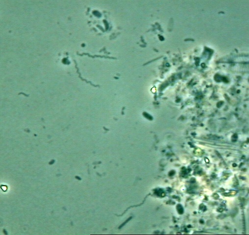
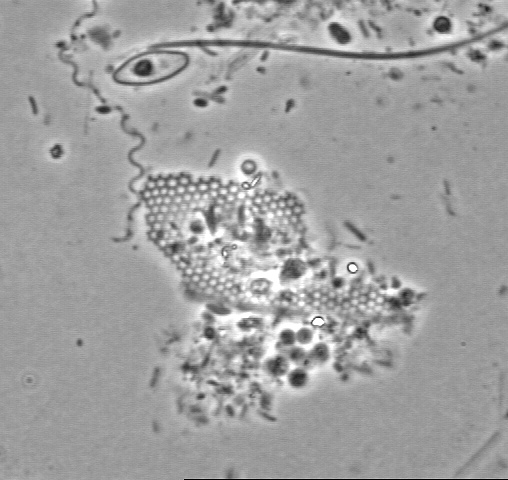
Black layer
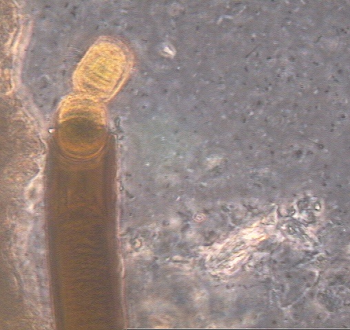
Microorganisms of the black, iron sulfide rich layer of the microbial mat from Great Sippewissett Saltmarsh, clustered around the remainings of an insect's eye (?). The spiral shaped organism (top left) is a Spirochaete.
Phase contrast micrograph
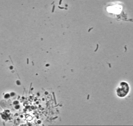
Black layer
The curved cells resemble typical sulfate reducing bacteria (Desulfovibrio spec).
Phase contrast, 75X75 µm
Black layer
Spirochaetes are easyly recognized by their irregular spiral shape. Living cells crawl between the sand particles; they show a typical twisting behaviour.
Phase contrast. 400x400 µm